Figure 2.

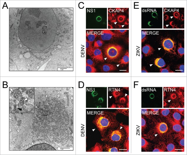
Flavivirus infection of HBMEC results in ER expansion. Transmission electron micrographs of (A) mock and (B) DENV (MOI of 3)-infected HBMEC 24 hpi. Black box shows area of zoomed-in region showing replication vesicles (white arrows) and immature virions (black arrows) within expanded ER. Scale bars: 2 µm. (C-F) Confocal microscopy of HBMEC infected with DENV (C and D) or ZIKV (E and F) at a MOI of 1; sites of ER expansion are marked with white arrowheads. Samples were stained 24 hpi for CKAP4, an ER sheet marker (red, C and E), and RTN4, an ER tubule marker (red, D and F). Sites of viral replication were determined by staining for DENV NS1 (green, C and D) or ZIKV dsRNA (green, E and F). Regions of colocalization are shown in yellow in merged images, with enhanced staining of ER markers shown at sites of viral replication. Scale bars: 20 µm.
