
Figure 2.
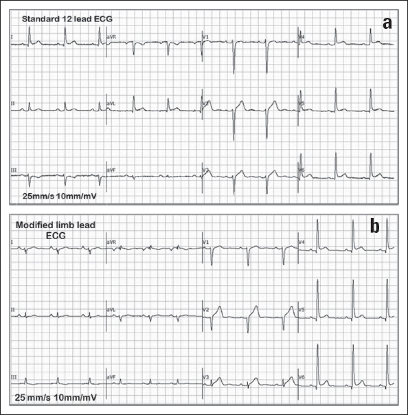
(a) Standard 12-lead ECG of a male subject in sinus rhythm. The R wave has maximum amplitude in all the leads. (b) MLL ECG of the same male subject in sinus rhythm used in the standard 12 lead ECG. The MLL ECG shows the presence of large P wave amplitudes and reduced R wave amplitudes in the limb leads