Introduction
Q fever is a zoonotic disease caused by Coxiella burnetii. The acute form of the disease may present as flu-like illness and pneumonia, whereas chronic form presents mainly as infective endocarditis (IE) (1).
Although acute cases and small epidemics of Q fever have been defined (2), herein we report the first case of chronic Q fever endocarditis in Turkey.
Case Report
A woman aged 29 years was admitted to hospital with symptoms of fever, weakness, and rash on her legs. She had undergone aortic surgery 3 times between 1997 and 2010: aortic commissurotomy because of rheumatic valve disease, aortic valve replacement due to aortic stenosis and probable IE, and aortic graft implantation and valve replacement because of aortic root dilatation, aneurysm, and pathologically confirmed blood culture negative IE. She was given 60 days of antibiotic therapy and discharged. She had fever, weakness, and weight loss afterwards. Patient had undergone transesophageal echocardiography (TEE) investigation 4 times, but none revealed findings compatible with endocarditis. Blood cultures were negative several times. She had recurrent nodular rash on her legs for 1½ years, which was diagnosed as leukocytoclastic vasculitis. A bloody sternal drainage had started after minor trauma to sternum 4 months before admission.
Patient frequently visited Yalova city of Turkey, site of last Q fever epidemic (published locally Kongre’s notes).
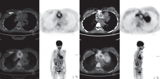
Patient was referred to hospital for further investigation of the vasculitis. Physical examination revealed discolored rash on her legs, 4/6 systolic murmur throughout cardiac foci, splenomegaly, and 1x1 cm open, draining wound on sternum. Transthoracic echocardiograms and TEE investigation were normal, and 3 sets of blood cultures were negative. Blood analyses were normal, with exception of the following: hemoglobin level: 8.1 gr/dL; leukocyte and platelet count 4400 cells/µL and 118 000 cells/µL, respectively; erythrocyte sedimentation rate (ESR) 94 mm/h; serum total protein, γ-globulin, C-reactive protein (CRP) and rheumatoid factor levels 8.9 g/dL, 3.34 g/dL, 13 mg/L, and 227 U/mL respectively; 1(+) cryoglobulin and 1(+) cryofibrinogen. She also had hematuria. Coxiella phase I IgG antibodies were positive at 1/262.144 titre. Positron emission tomography–computed tomography (PET/CT) revealed fluorodeoxyglucose uptake around aortic valve and graft, and in mediastinum and sternum (Fig.1). Patient was diagnosed with IE, aortic graft infection, mediastinitis, and sternal osteomyelitis due to C. burnetii. Doxycycline 2x100 mg/day and hydroxycloroquine 3x200 mg/day orally, and ciprofloxacin 2x400 mg/day intravenously were initiated. She underwent aortic valve and graft replacement. All intraoperative tissue samples (valve, graft, mediastinum, sternum) were positive for C. burnetii with PCR. Patient died as result of perioperative cerebellar infarct.
Figure 1.
Positron emission tomography–computed tomography images of the patient. Black arrows indicate fluorodeoxyglucose uptake around aortic valve, aortic graft, and in mediastinum and sternum
Discussion
Patients with underlying cardiac valvular or vascular disease have increased risk of chronic Q fever (1). Present patient was the first case of chronic Q fever endocarditis from Turkey. She probably acquired the infection in Yalova city.
Major presentations of chronic Q fever infection, endocarditis, vascular graft infection, and osteomyelitis were all present; mediastinitis and sternal osteomyelitis probably developed as progression of untreated aortic graft infection over the years. There is 1 other case report of sternal osteomyelitis and aortitis in patient who underwent prosthetic aortic valve and graft replacement. That patient also had very subtle progression and infection that was diagnosed 4 years after first signs (3). Especially in countries where the disease is not known, such as Turkey, diagnosis may be delayed for years. For current patient, despite detailed diagnostic evaluations over 5 years, diagnosis of chronic Q fever was not made.
Other reasons for diagnostic delay in chronic Q fever endocarditis were indolent clinical presentation, absence of vegetation in echocardiographic findings, blood culture negativity, and misdiagnosis due to presence of autoantibodies and immune complexes (1, 4–7).
Microbiological diagnosis of chronic Q fever mainly relies on serology and Coxiella phase-I IgG titre ≥1:800, which is a major Duke criterion (1). Coxiella phase-I Ig G titre was found to be 1/262.144 in present patient, which is unusually high compared to previous reports (5, 7, 8), probably because of extensive infection due to delayed diagnosis.
Diagnostic delay has a significant impact on patient’s prognosis and risk of complications. Physician’s experience with the disease can reduce delay (9), and lack of familiarity probably contributed to outcome of this patient.
PETscan showing specific valve fixation is also a major criterion for Q fever endocarditis (1) and contributed to diagnosis in present patient.
Conclusion
Although not reported before in Turkey, chronic Q fever endocarditis should be suspected in patients with known valvulopathy and unexplained prolonged fever, purpuric skin rash, persistent hepatosplenomegaly, increased ESR, CRP or autoantibody level, cryoglobulinemia, or necessity for early valve replacement after cardiac surgery, even in the absence of vegetation on echocardiography or positive blood cultures.
References
- 1.Million M, Raoult D. Recent advances in the study of Q fever epidemiology, diagnosis and management. J Infect. 2015;71:S2–S9. doi: 10.1016/j.jinf.2015.04.024. [DOI] [PubMed] [Google Scholar]
- 2.Yıldırmak T, Şimşek F, Çelebi B, Çavuş E, Kantürk A, Efe-İris N. A rare case of acute Q fever presenting with deep jaundice and a review of the literature. Klimik Derg. 2010;23:124–9. [Google Scholar]
- 3.Ghassemi M, Agger WA, Vanscoy RE, Howe GB. Chronic sternal wound infection and endocarditis with Coxiella burnetii. Clin Infect Dis. 1999;28:1249–51. doi: 10.1086/514797. [DOI] [PubMed] [Google Scholar]
- 4.Million M, Thuny F, Richet H, Raoult D. Long-term outcome of Q fever endocarditis: a 26-year personal survey. Lancet Infect Dis. 2010;10:527–35. doi: 10.1016/S1473-3099(10)70135-3. [DOI] [PubMed] [Google Scholar]
- 5.Raoult D, Tissot-Dupont H, Foucault C, Gouvernet J, Fournier PE, Bernit E, et al. Q Fever 1985-1998. Clinical and epidemiologic features of 1,383 Infections. Medicine (Baltimore) 2000;79:109–23. doi: 10.1097/00005792-200003000-00005. [DOI] [PubMed] [Google Scholar]
- 6.Lefebvre M, Grossi O, Agard C, Perret C, Le Pape P, Raoult D, et al. Systemic immune presentations of Coxiella burnetii infection (Q fever) Semin Arthritis Rheum. 39:405–9. doi: 10.1016/j.semarthrit.2008.10.004. [DOI] [PubMed] [Google Scholar]
- 7.van der Hoek W, Versteeg B, Meekelenkamp JCE, Renders NHM, Leenders ACAP, Weers-Pothoff I, et al. Follow-up of 686 Patients with acute Q fever and detection of chronic infection. Clin Infect Dis. 2011;52:1431–6. doi: 10.1093/cid/cir234. [DOI] [PubMed] [Google Scholar]
- 8.Gunn TM, Raz GM, Turek JW, Farivar RS. Cardiac manifestations of Q fever infection: Case series and a review of the literature. J Card Surg. 2013;28:233–7. doi: 10.1111/jocs.12098. [DOI] [PubMed] [Google Scholar]
- 9.Houpikian P, Habib G, Mesana T, Raoult D. Changing clinical presentation of Q fever endocarditis. Clin Infect Dis. 2002;34:e28–31. doi: 10.1086/338873. [DOI] [PubMed] [Google Scholar]


