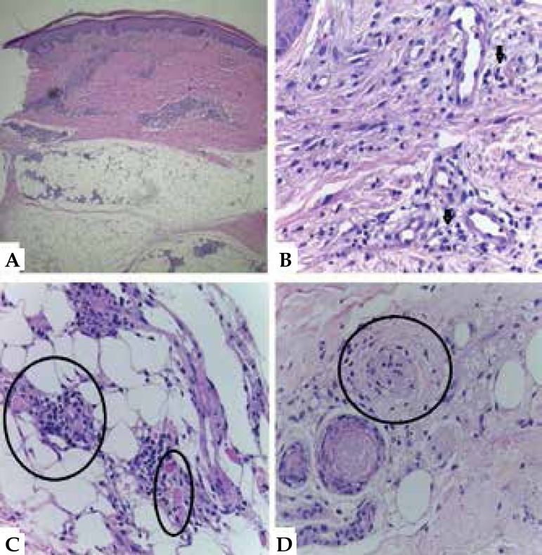
Figure 3.
A) Inflammatory infiltrate affecting the dermis and hypodermis (Hematoxylin - eosin, x40). B) Perivascular lymphohistiocytic infiltrate with multiple Virchow’s cells (see arrows)(Hematoxylin - eosin, x100). C) Thrombosis of vessels in the hypodermis (see circles)(Hematoxylin - eosin, x100). D) Endothelial proliferation with occlusion of the vessel lumen (see circle), (Hematoxylin - eosin, x100)

