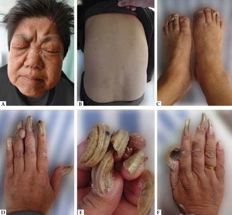
Figure 1.
A: Extensive, smooth, skin-colored, papules involving the malar region in a butterfly distribution. B: A hypomelanotic macule and several plaques with variable size and shape located on her back. C-F: Multiple fusiform, strawberry-like and vermiform, periungual reddish tumors extended from the nail groove. Underlying nail plate distorted, deformed, and protracted resembling a ram’s horn

