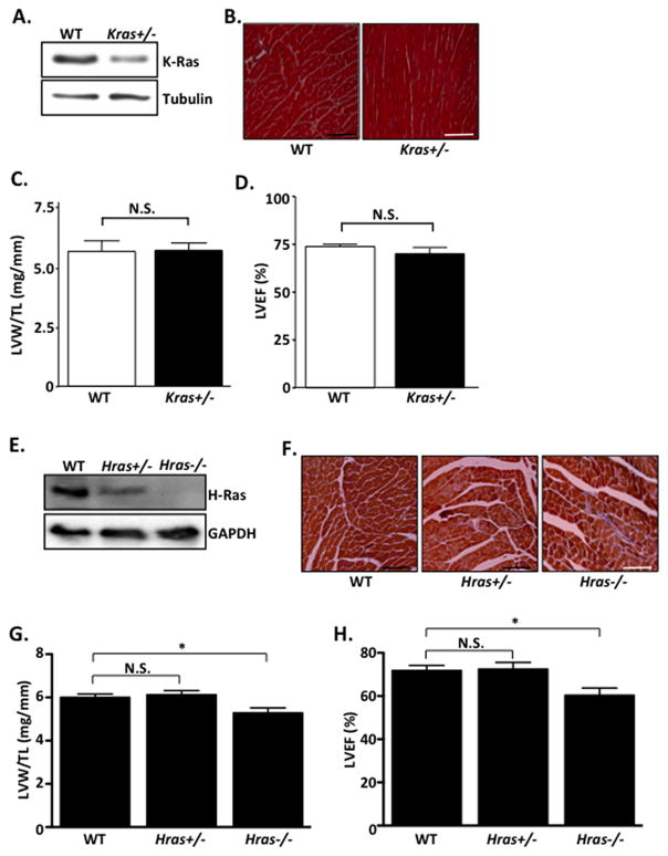
Figure 1. Basal cardiac characterization of K- and H-Ras mutant mice.
A–H. All mice were between 10–12 weeks old. A. Ventricular homogenates from WT and Kras+/− mice were subjected to western blot. B. Heart sections were stained with Masson’s Trichrome to visualize interstitial fibrosis. C. Post-mortem gravimetric analysis of left ventricular weight/tibia length (LVW/TL) was determined. D. Prior to sacrifice, echocardiographic analysis was performed and left ventricular ejection fraction (%LVEF) determined. N=5 per group. E. Ventricular homogenates from WT, Hras+/− and Hras−/− mice were subjected to western blot. F. Heart sections were stained with Masson’s Trichrome to visualize interstitial fibrosis. G. Post-mortem gravimetric analysis of left ventricular weight/tibia length (LVW/TL) was determined. H. Prior to sacrifice, echocardiographic analysis was performed and left ventricular ejection fraction (%LVEF) determined. N=6–10 per group. Data are mean ± SEM. *,P<0.05. N.S.=not significant.

