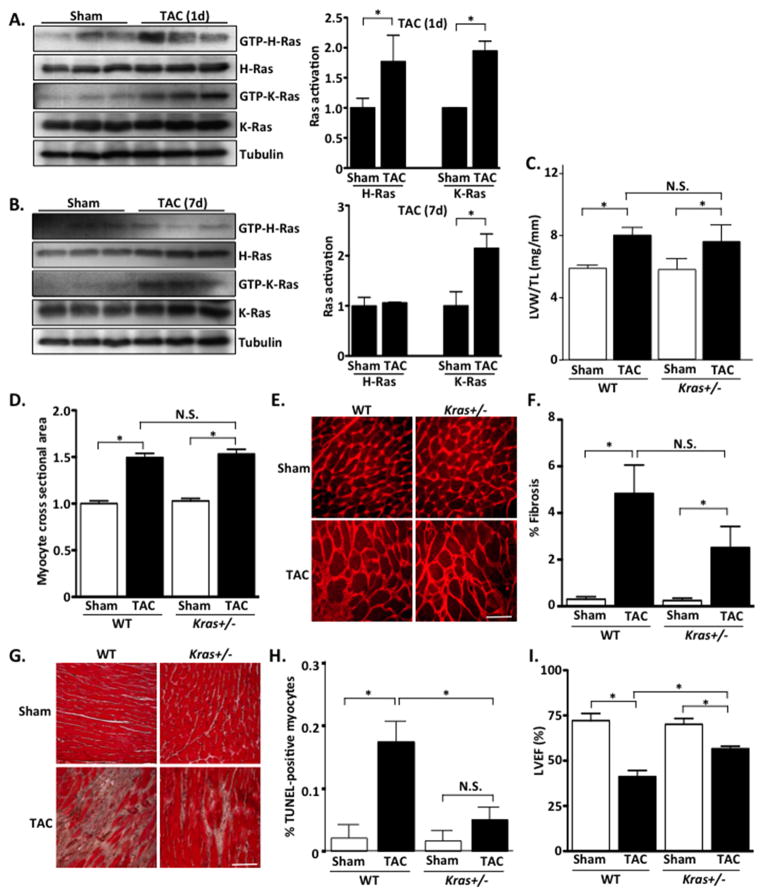
Figure 2. Inhibition of endogenous K-Ras does not alter hypertrophy but protects against pressure-overload-induced cardiac dysfunction.
A and B. WT mice were subjected to TAC or sham operation for 1 or 7 days. Ventricular homogenates were subjected to RBD-pull down assay to determine activation of H- and K-Ras. Quantification of blots is shown in right panels. C. Left ventricular weight/tibia length (LVW/TL) was determined in TAC and sham operated groups. D. Cardiomyocyte cross-sectional area was determined by wheat germ agglutinin (WGA) staining. E. Representative WGA images. F. Fibrosis was determined by Masson’s Trichrome staining. G. Representative images shown. Scale bar, 100 μm. H. Extent of apoptosis was determined by TUNEL staining. I. Echocardiographic analysis was performed to determine left ventricular ejection fraction (%LVEF). N=4–9 per group. Data are mean ± SEM. *,P<0.05. N.S.=not significant.

