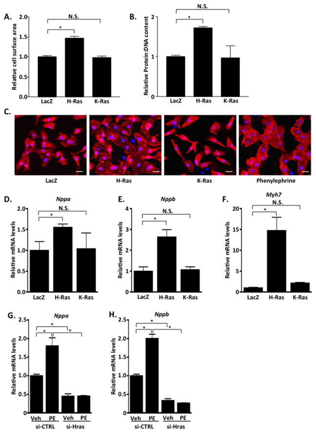
Figure 4. H-Ras, but not K-Ras, promotes cardiomyocyte hypertrophy in vitro.
A–F. neonatal rat cardiomyocytes (NRCMs) were treated with LacZ, H-Ras12V or K-Ras12V adenovirus and assayed 48 hours later. A. NRCMs were stained with troponin T to visualize cardiomyocytes and cell surface area determined. B. Cells were collected and protein and DNA concentrations were determined. C. Representative images shown. Scale bar, 30 μm. mRNA was isolated and qRT-PCR performed to determine Nppa (D), Nppb (E) and Myh7 (F) levels. NRCMs were treated with siRNA to deplete endogenous H-Ras (si-Hras) or control siRNA (si-CTRL). 72 hours later, cells were treated with phenylephrine (100μM; 24 hours) or vehicle and qRT-PCR performed to determine Nppa (G) and Nppb (H) levels. N=3. Data are mean ± SEM. *,P<0.05. N.S.=not significant.

