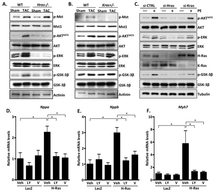
Figure 5. H-Ras activates the PI3K-AKT signaling axis and promotes cardiomyocyte hypertrophy.
A and B. Ventricular homogenates from control and mutant mice were subjected to western blot analysis to examine downstream signaling in response to 4 week TAC. C. NRCMs were treated with control siRNA (si-CTRL), or siRNA targeted to H-Ras (si-Hras) or K-Ras (si-Kras) for 72 hours. Cells were then stimulated with phenylephrine (PE; 100 μM) or vehicle control for 1 hour. NRCMs were transduced with LacZ or H-Ras12V in combination with vehicle or inhibitor for 48 hours and qRT-PCR performed to detect Nppa (D), Nppb (E) and Myh7 (F). N=3–4. Data are mean ± SEM. *,P<0.05.

