Fig. 2.
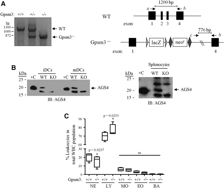
Loss of AGS4 results in altered leukocyte population phenotype. (A) Left panel: A three-primer PCR approach was used to genotype AGS4/Gpsm3 wild-type (+/+), heterozygous (+/−) and null (−/−) mice. Right panel: Schematic depicting the strategy used to generate and polymerase chain reaction (PCR) genotype AGS4/Gpsm3-null mice as described in Materials and Methods. DNA primers a, b, and c (corresponding to “Gpsm3 16651 forward,” “Common 3’ forward” and “CSD-Gpsm3-SR1,” respectively; see Materials and Methods for additional details) were used in a three-primer PCR reaction in which a wild-type product at 1200 bp resulted from priming from primers a and b, and an AGS4/Gpsm3−/− product at 776 bp resulted from priming from primers b and c. (B) Lysates (100 μg/lane) from primary immature dendritic cells (iDCs), mature dendritic cells (mDCs) (left panel), and splenocytes (right panel) from WT (wild-type), and Gpsm3-null mice were subjected to SDS-PAGE, transferred to PVDF membranes, and immunoblotted (IB) with AGS4 antisera as described in Materials and Methods. “+C” refers to lysate prepared from HEK293 cells transfected with pcDNA3::AGS4 after 24 hours as described in Materials and Methods. (C) Complete blood count (CBC) analysis was from blood collected from WT and AGS4/Gpsm3-null mice as described in Materials and Methods. The percentage of leukocyte populations in relation to total number of white blood cells was calculated and compared between WT and AGS4/Gpsm3-null littermate pairs. BA, basophils; EO, eosinophils; LY, lymphocytes; MO, monocytes; NE, neutrophils. Data are represented as the mean ±S.E. from four pairs of 12-week-old mice.

