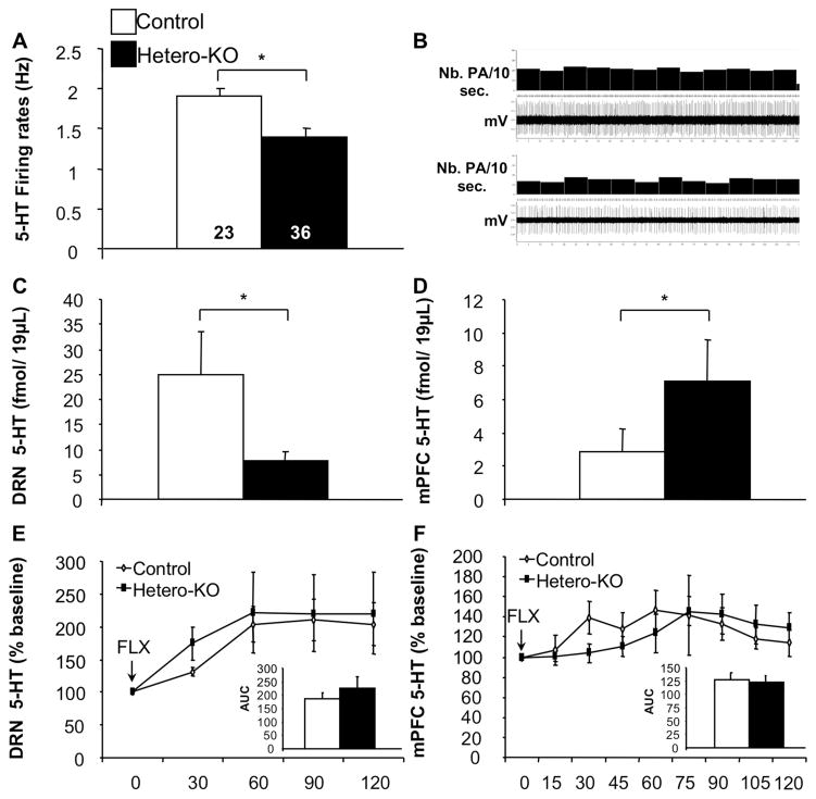
Figure 3. Suppression of 5-HT1A heteroreceptors results in altered 5-HT tone.
(A) Hetero-KO mice displayed lower basal 5-HT firing rates compared to controls (ANOVA for main effect of group: F(1,57)=4.647; p<0.05; n=23–36/group). (B) Representative recordings of DRN 5-HT neurons obtained in control (Top) and Hetero-KO (Bottom). (C) Decreased basal 5-HT levels in the DRN in Hetero-KO mice (ANOVA for main effect of group: F(1,14)=4.865; p<0.05)(D) along with increased basal 5-HT levels in the mPFC in Hetero-KO mice (ANOVA for main effect of group: F(1,14)=4.629; p<0.05). (E) Area under the curve analysis, revealed no significant effect of group in the DRN (ANOVA for main effect of group: F(1,14)=0.144, p=0.708) but a significant effect of time (ANOVA repeated measures for main effect of time: DRN: F(1,14)=6.215, p<0.01) in 5-HT levels following injection of FLX. (F) 5-HT measured by area under the curve analysis, revealed no significant effect of group in the mPFC (ANOVA for main effect of group: F(1,14)=0.432, p=0.521) but a significant effect of time (ANOVA repeated measures for main effect of time: F(1,14)=3.387, p<0.01) measured after FLX. (n=7–9/group). Data are represented as mean±SEM. *p≤0.05, **p<0.01.

