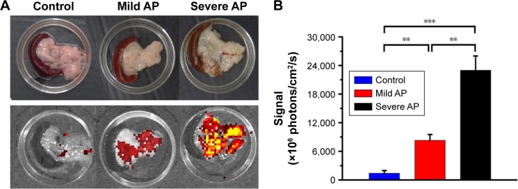
Figure 6.
The in vivo pancreas distribution of DiR-loaded M-Gd-NL.
Notes: After the mild or severe AP SD rat models were established, the rats were treated with M-DiR-NL. M-DiR-NL was injected intravenously as a single dose (500 μg DiR/kg) via tail vein. The rats were anesthetized by inhalation. Four hours after injection, the rats were euthanized and the excised pancreas was imaged with IVIS® Lumina II Imaging System and recorded by a built-in CCD camera. (A) A representative excised pancreas was shown. (B) Quantitative fluorescence analysis of excised pancreases. All comparisons were performed between the two groups by one-way analysis of variance with Newman–Keuls posttest. Data are expressed as mean ± standard deviation (n=3). **P<0.01; ***P<0.001.
Abbreviations: AP, acute pancreatitis; DiR, 1,1′-dioctadecyl-3,3,3′,3′-tetramethylindotricarbocyanine iodide; M-DiR-NL, mannosylated DiR-loaded liposomes; M-Gd-NL, gadolinium-diethylenetriaminepentaacetic-loaded mannosylated liposomes; SD, Sprague Dawley.

