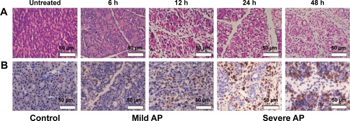
Figure 7.
The demonstration of AP model by HE staining, and the macrophages in AP are revealed by CD68 staining.
Notes: After the establishment of AP model (mild AP was established 6 or 12 h by three IP injections of L-arginine, and severe AP was established 24 or 48 h by three IP injections of L-arginine), the pancreases were rapidly dissected and washed with 0.9% physiological saline and fixed by immersion in 4% paraformaldehyde. (A) HE staining was performed to observe whether the experimental AP model was established. (B) Immunohistochemistry was performed to observe the macrophage infiltration in the pancreas by CD68 staining. The macrophages in the pancreas were revealed by using mouse anti-rabbit CD68 antibody (Abcam) as the primary antibody and goat anti-mouse IgG antibody (horseradish peroxidase [HRP] labeled) as the secondary antibody.
Abbreviations: AP, acute pancreatitis; HE, hematoxylin–eosin.

