Figure 9.
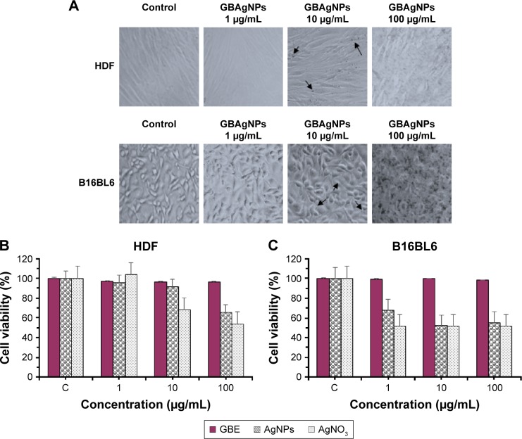
Comparative study of the effect of GBAgNPs, GBE and AgNO3 on cell viability and on the morphology of HDF and B16 cells.
Notes: Optical microscopy images (A) of HDF and B16 cell lines (40× magnification) at 72 h after treated with 1–100 μg/mL of GBAgNPs. Treated HDF and B16 cells with silver (10 μg/mL) could be identified by dark dense clusters which are indicated with arrows; no cluster was found in control groups. Cytotoxicity effect of GBE, GBAgNPs, and silver salts on HDF (B) and B16 (C) cell lines; cell viability decreased with an increase in the concentration of GBAgNPs. Cell viability was determined by MTT assay. Data are expressed as a percentage of sample-treated control and presented as mean ± SEM of three separate experiments.
Abbreviations: B16, murine melanoma B16Bl6 cells; GBAgNPs, silver nanoparticles from ginseng berry; GBE, ginseng berry extract; HDF, human dermal fibroblast; MTT, 3-(4,5-dimethyl-thiazol-2yl)-2, 5-diphenyl tetrazolium bromide; SEM, standard error of the mean.

