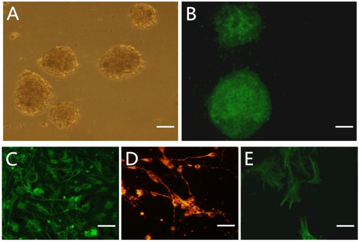
Fig 1. Characterization and differentiation of NSCs.
A: phase-contrast image of NSCs globes cultured 5d in NSCs culture medium. B: Immunostaining of NSCs with Nestin antibody. C-F: immunostaining of differentiated cells with astrocyte marker GFAP, neuron marker Tuj-1 and oligodendrocyte marker O4 in 10% FBS-DF12 for 5 days. Scale bar: A-B 400 um; C-F 20 um.

