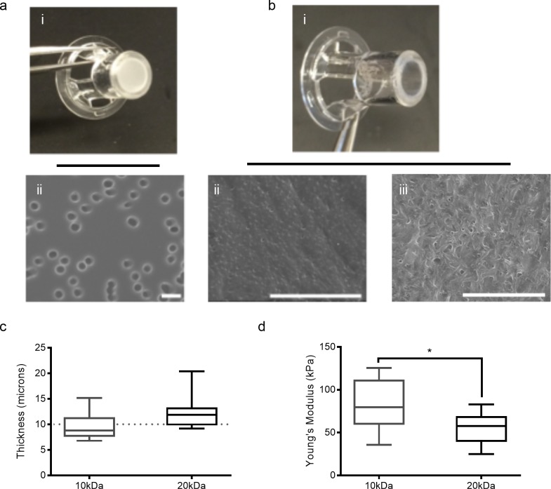
Fig 1. Zinc oxide micro-needles introduce pores into PEG hydrogels.
(a-b) Images of commercial TWs (a-i) and porated PEG hydrogels in TW casing (b-i). SEM micrographs of TWs (a-ii), 10kDa (b-ii), and 20kDa (b-iii) porated PEG hydrogels; scale bars: 5 microns. (c) Hydrogel thickness as determined by OCT. Whiskers denote minimum and maximum value; there is no statistical difference between 10kDa and 20kDa hydrogels. (d) Young’s modulus of 10kDa and 20kDa porated PEG hydrogels. Whiskers denote minimum and maximum value; *p<0.05.

