FIGURE 2.

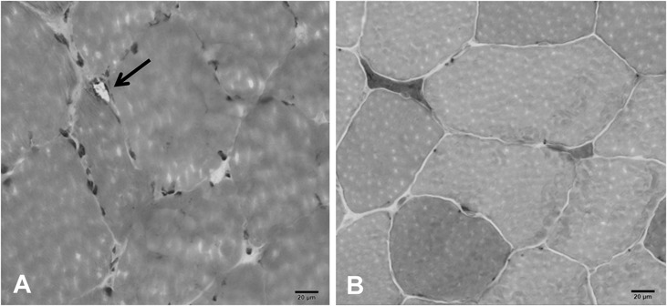
Left biceps brachii muscle biopsy. A, High power hematoxylin and eosin stain revealing a small peripheral vacuole in a nonnecrotic fiber as noted with arrow. B, Atrophic muscle fibers overreacting for nonspecific esterase.

Left biceps brachii muscle biopsy. A, High power hematoxylin and eosin stain revealing a small peripheral vacuole in a nonnecrotic fiber as noted with arrow. B, Atrophic muscle fibers overreacting for nonspecific esterase.