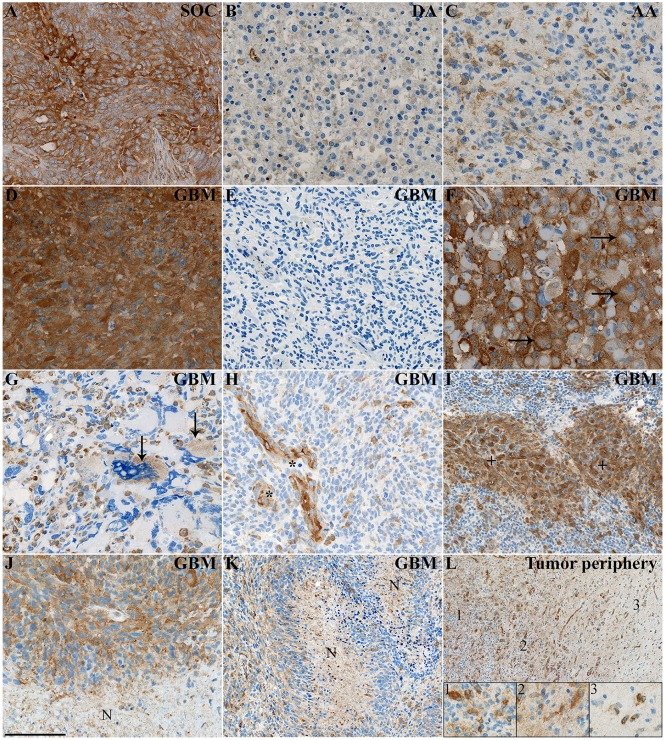
Fig 2. Immunohistochemical stainings of MMP-2.
(A) MMP-2 showed intense expression in the serous ovarian carcinoma (SOC) which was used as a positive control. (B-C) MMP-2 was weakly expressed in diffuse astrocytomas (DA), while it exhibited stronger expression levels in anaplastic astrocytomas (AA). (D) The majority of glioblastomas (GBMs) had areas with intense MMP-2 expression. (E) However, a few GBMs were mostly MMP-2 negative. (F) Gemistocytic tumor cells often expressed high levels of MMP-2, especially in the membrane (horizontal arrows). (G) Multinucleated giant cells expressed limited amounts of MMP-2 (vertical arrows). (H) Blood vessels (asterisks) including (I) glomeruloid blood vessels (plus signs) were largely MMP-2 positive and often exhibited intense staining. (J-K) Cells surrounding larger necrotic areas (N) often showed intense MMP-2 positivity, while the intensity appeared lower in areas with pseudopalisading necroses (N). (L) MMP-2 positive cells were present in higher amounts in the central tumor (insert 1), but some intensely stained cells were also found in the border zone (insert 2) as well as in the tumor periphery (insert 3), where there was a considerably lower cellularity. Scale bar 100 μm (A-H, J), 400 μm (I, K, L).

