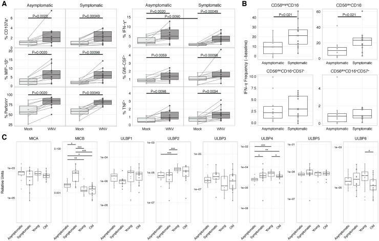
Fig 5. Robust functional response to WNV by NK cells from asymptomatic and symptomatic WNV subjects.
PBMCs from asymptomatic (n = 10) and symptomatic (n = 12) WNV subjects were incubated with medium alone (mock) or infected with WNV (MOI = 1) for 24 h. (A) Total NK cells from mock and WNV-infected samples of each WNV subject were compared by mass cytometry for surface expression of CD107a and production of MIP-1β, perforin, IFN-γ, TNF, and GM-CSF. Mock-WNV comparisons, paired Wilcoxon tests. Mock-mock and WNV-WNV comparisons, Mann-Whitney U tests. (B) Four NK cell subsets from asymptomatic and symptomatic WNV subjects were compared for induction of IFN-γ by subtracting baseline of the mock group from the WNV-infected group. Mann-Whitney U tests. (C) Samples from all four groups of subjects (n = 56) were compared by qPCR for baseline expression of MICA, MICB, ULBP1, ULBP2, ULBP3, ULBP4, ULBP5, and ULBP6. Mann-Whitney U tests. P < 0.05 = *; P < 0.01 = **; P < 0.001 = ***.

