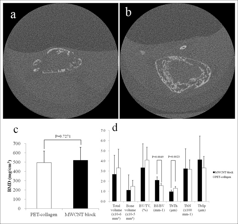
Fig 6. The μCT evaluation at 3 weeks after implanting rhBMP-2-containing scaffold in the mouse back muscle.
(a) CT image of ectopic bone formed using a PET-fiber-reinforced collagen sheet (positive control) as a scaffold (arrow). (b) CT image of ectopic bone formed using MWCNT blocks. Distinct bone formation was observed in both cases. (c) BMD values of ectopic bone formed around an rhBMP-2-containing PET-fiber-reinforced collagen sheet and rhBMP-2-containing MWCNT block. (d) Three-dimensional μCT analysis. BV/TV: bone volume / total volume; BS/BV: bone surface / bone volume; TbTh: trabecular thickness; TbN: trabecular number; TbSp: trabecular spacing.

