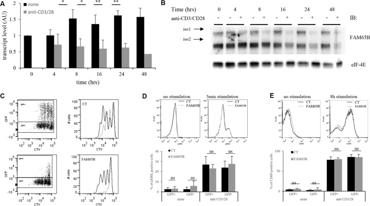
Figure 4. FAM65B expression is inversely correlated with the proliferative potential of activated T cells.
Primary human T cells were stimulated in vitro for several hours using anti-CD3 and -CD28 coated-beads. FAM65B expression was quantified at both the transcript (A) and the protein (B) levels. (C) Primary human T cells were transfected with GFP (Control: CT) or FAM65B-GFP expression vector then stained with Cell Trace Violet (CTV) and stimulated in vitro as described in (A) for 4 days. In the histograms, the grey lines correspond to the GFP+ cells and the black lines to the GFP− cells. Note that in CT conditions, GFP+ cells proliferate slightly less efficiently than GFP− cells. This is likely to be an effect of the transfection per se. (D) ERK phosphorylation and (E) CD69 expression of transfected cells stimulated or not with anti-CD3 and –CD28 was analyzed by flow cytometry after 3 minutes and 8 hours of culture, respectively.

