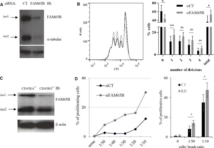
Figure 5. FAM65B expression is inversely correlated with the T cells activation threshold.
(A) The efficiency of FAM65B knock-down in human primary T cells was checked by immunoblotting with anti-FAM65B and anti-α-tubulin as a loading control. (B) Human primary T cells were transfected with control (siCT) or FAM65B (siFAM65B) siRNA, then stained with CTV and stimulated in vitro for 3 days using anti-CD3 and -CD28 coated-beads. The proliferation of transfected cells was analyzed by flow cytometry after 3 days of stimulation. The percentage of cells exhibiting 0, 1, 2, 3, 4 divisions or the addition of them (total) is quantified as the mean ± SD obtained from four donors. (C) Naive (CD45RA+) or activated/memory (CD45RO+) populations were purified from peripheral blood T cells. FAM65B expression was then analyzed by immunoblotting. (D) Primary human CD45RA+ T cells transfected with siFAM65B or siCT were stained with CTV and stimulated in vitro using variable doses of anti-CD3 and anti-CD28 -coated beads. The proliferation of the transfected cells was analyzed by flow cytometry after 3 days of stimulation. Results are representative of at least three individual experiments

