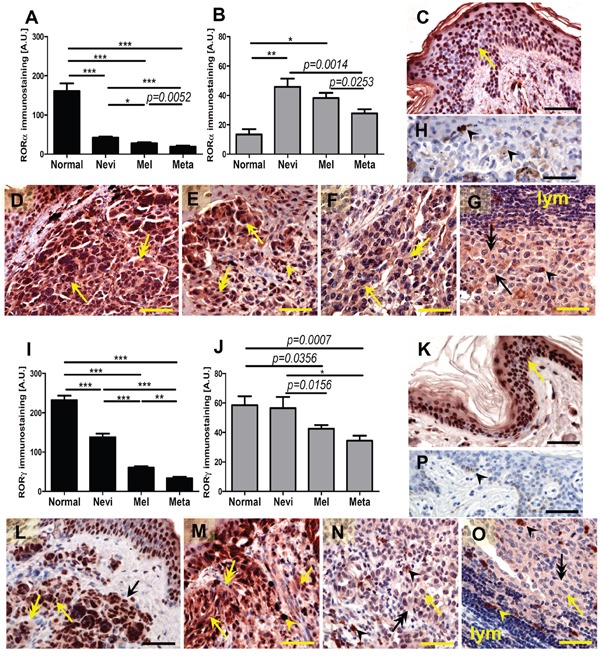
Figure 1. The mean level of nuclear (A, I) and cytoplasmic (B, J) RORα (A, B) and RORγ (I, J) in keratinocytes of normal skin, melanocytic cells of nevi, primary melanomas (MM) and metastases (meta).

Statistically significant differences are denoted with P values as determined by Student's t-test and with asterisks by ANOVA (*P<0.05, **P<0.01 and ***P<0.001). Representative RORα (C-H) and RORγ (K-P) immunostaining of normal skin (C, K), melanocytic nevi (D, L), early-stage melanomas (E, M), advanced melanomas (F, N) and lymph node metastases (G, O) Negative controls are presented at H and P panels. Arrows indicate nuclear staining in melanomas, arrow heads - melanin, double arrows - cytoplasmic staining, lymph - lymphocytes, scale bars - 50μm.
