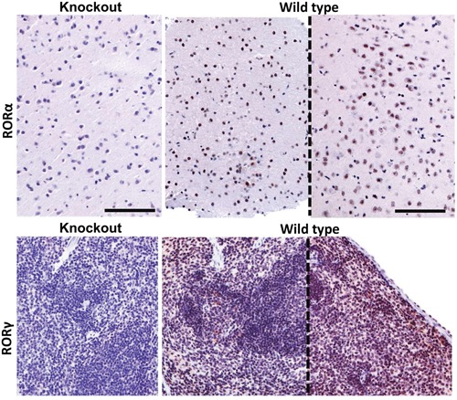
Figure 14. RORα (upper panel) and RORγ (lower panel) immunostaining of brain and spleen tissue sections of RORα or RORγ knockout mice and wild type mice (sections from two different mice, separated with dashed line).

The mouse brain samples were stained with anti-RORα antibody and mouse spleen with anti-RORγ antibody. Scale bars - 100μm.
