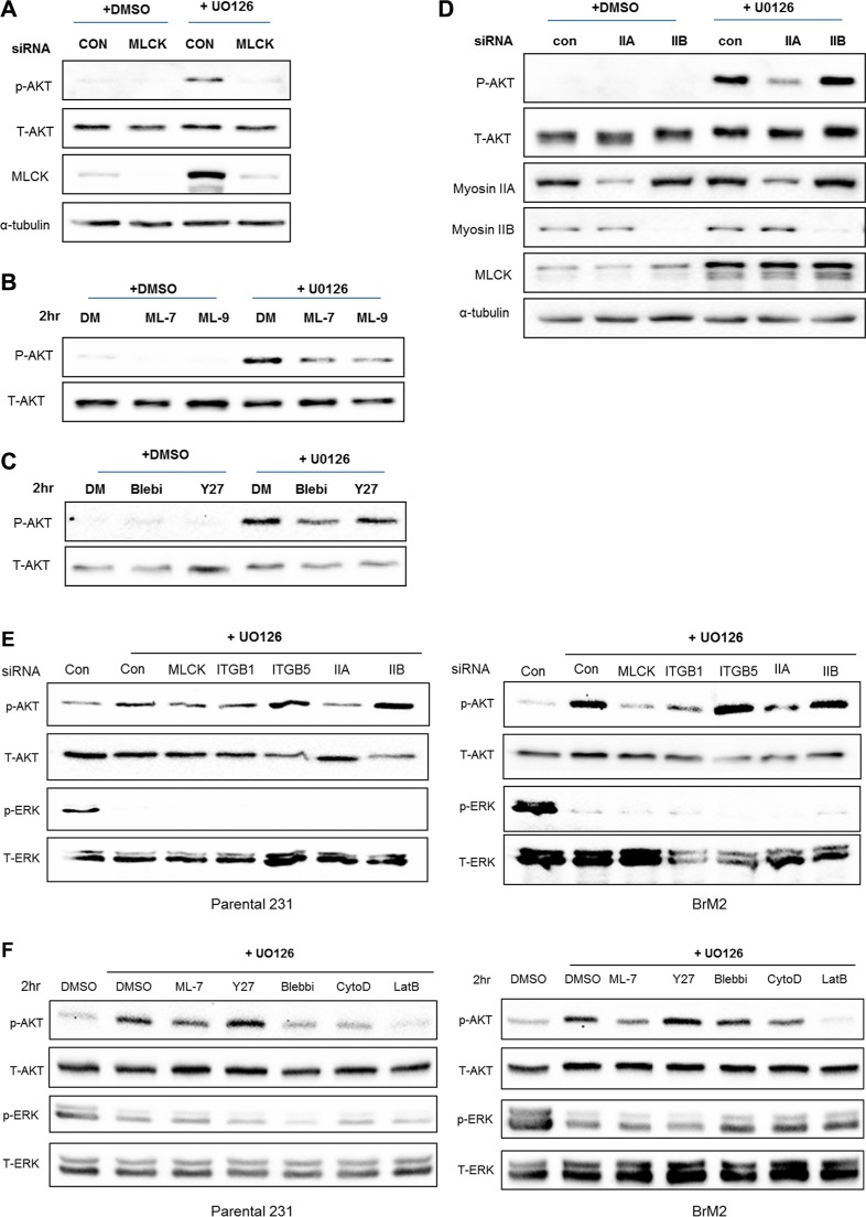
Figure 7. MLCK and Myosin IIA are required for PI3K-AKT activation following MEK suppression.
(A) Control siRNA or MLCK siRNA were transfected in LM2 cells prior to treating with either DMSO or UO126 (25 μM). After an additional 48 hr incubation of drug treatment cells were collected with 2× lamelli sample buffer and immunoblotted with antibodies against phosphorylated AKT, AKT, MLCK. α-tubulin was used for protein loading control. (B) LM2 cells were treated with pharmacological inhibitors of MLCK, ML-7 and ML-9 respectively pre-incubated in either DMSO or 25 μM UO126 for 48 hr. (C) LM2 cells were acutely treated with Blebbistatin (50 μM) or Y27632 (20 μM) for 2 hr, which is pharmacological inhibitors of myosin II and ROCK respectively pre-incubated in either DMSO or 25 μM UO126 for 48 hr. (D) LM2 cells were transfected with either control siRNA or specific myosin II isoform, IIA and IIB respectively. After transfected, cells were incubated with DMSO or 25 μM UO126 for an additional 48 hr. (E) Prior to western blot analysis, Parental and BrM2 were pre-treated with UO126 for 48 hr. Then, cells were treated with various actin cytoskeleton disrupting drugs including ML-7 (20 uM), Y27632 (10 uM), Blebbistatin (50 uM), Cytocalasin D (1 uM) and Latruculin B (5 uM) for 2 hr. (F) Parental and BrM2 cells were transfected with individual siRNA against MLCK, integrin β1, integrin β5, myosin IIA and myosin IIB. After then Cells were treated with UO126 for 48 hr. AKT activation was measured by immunoblotting with phosphorylated AKT antibody detecting S473 phosphorylation.

