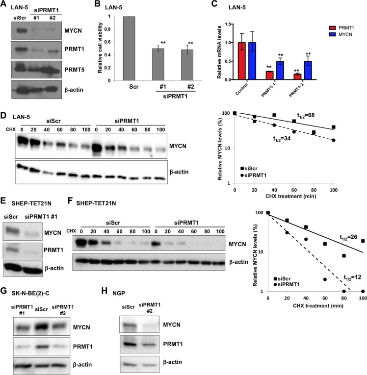
Figure 3. PRMT1 regulates MYCN expression.
(A) Western blot analysis of PRMT1 knockdown in LAN-5 cells 6 days following transfection with two independent PRMT1 siRNAs with indicated antibodies. β-actin was used as a loading control. (B) PRMT1 knockdown reduces cell viability. Cell viability was measured using the Trypan blue viability test and results are expressed as percentage of viable cells. Mean ± SEM (n = 3, Student's t test, **P < 0.01). (C) RT-qPCR analysis of PRMT1 and MYCN mRNA levels in siPRMT1-transfectd LAN-5 cells. Mean ± SEM (n = 3, Student's t test, **P < 0.01). (D) Immunoblots with anti-MYCN and β-actin (loading control) after CHX treatment following transient transfection of scrambled siRNA (siScr) or siPRMT1#1 in LAN-5 cells. Plots of densitometric quantification of MYCN protein stability were shown. (E) Western blot analysis of PRMT1 knockdown in SHEP-TET21N cells (Tet-off, MYCN on) 6 days following transfection with siPRMT1 #1 with indicated antibodies. β-actin was used as a loading control. (F) Immunoblots with anti-MYCN and β-actin after CHX treatment following transient transfection of scrambled siRNA (siScr) or siPRMT1#1 in SHEP-TET21N cells (Tet-off, MYCN on). Plots of densitometric quantification of MYCN protein stability were shown. Western blot analysis of PRMT1 knockdown in SK-N-BE(2)-C (G) and NGP cells (H) 6 days following transfection with PRMT1 siRNAs.

