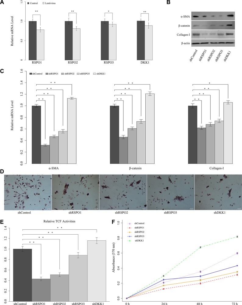
Figure 4. Knockdown of RSPOs (RSPO1, RSPO2, and RSPO3) repressed WNT pathway activity and HSCs activation, whereas knockdown of DKK1 enhanced RSPOs' expression.
(A) Real-time PCR results showed that lentivirus delivery decreased the mRNA level of both RSPOs and DKK1. (B) Knockdown of RSPOs down-regulated the expression of α-SMA, nuclear β-catenin, and Collagen-I compared with the control, whereas knockdown of DKK1 enhanced the expression of α-SMA, nuclear β-catenin, and Collagen-I. (C) Real-time PCR results were consistent with the findings of Western blot assay. (D) Oil Red O staining showed that knockdown of RSPOs increased the lipid droplets in HSCs, while knockdown of DKK1 reduced the lipid droplets (bar = 50 μm, magnification × 400). (E) TCF activities significantly decreased in RSPOs-knockdown HSCs compared with the control, while DKK1-knockdown significantly increased TCF activities. (F) MTT assay showed RSPOs-knockdown suppressed HSCs proliferation, whereas DKK1-knockdown promoted HSCs proliferation. Data represents the mean of three independent experiments, and error bars are standard deviation of means. *p < 0.05 compared with the control (CCl4-induced hepatic fibrosis mice transfected with scrambled siRNA through Lentivirus transduction), **p < 0.01 compared with the control.

