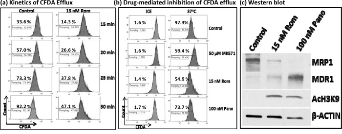
Figure 3. Functional assay of MRP1 protein.

PEER cells were exposed to solvent (Control) or 15 nM romidepsin (Rom) for 48 hrs, harvested, incubated with the MRP1 substrate CFDA for 20 min, shifted to 37°C, and analyzed for efflux of CFDA at the indicated time point by flow cytometry a. To determine drug-mediated inhibition of CFDA efflux, cells were exposed to 15 nM Rom or 100 nM Pano (panobinostat) for 48 hrs or to the MRP1 inhibitor MK571 (50 μM) for 30 min prior to the efflux assay. CFDA efflux was performed for 30 min b. “Count” refers to the number of live (i.e., PI-negative) cells. Western blot analysis confirmed the 48-hr drug-mediated decrease in MRP1 protein level c.
