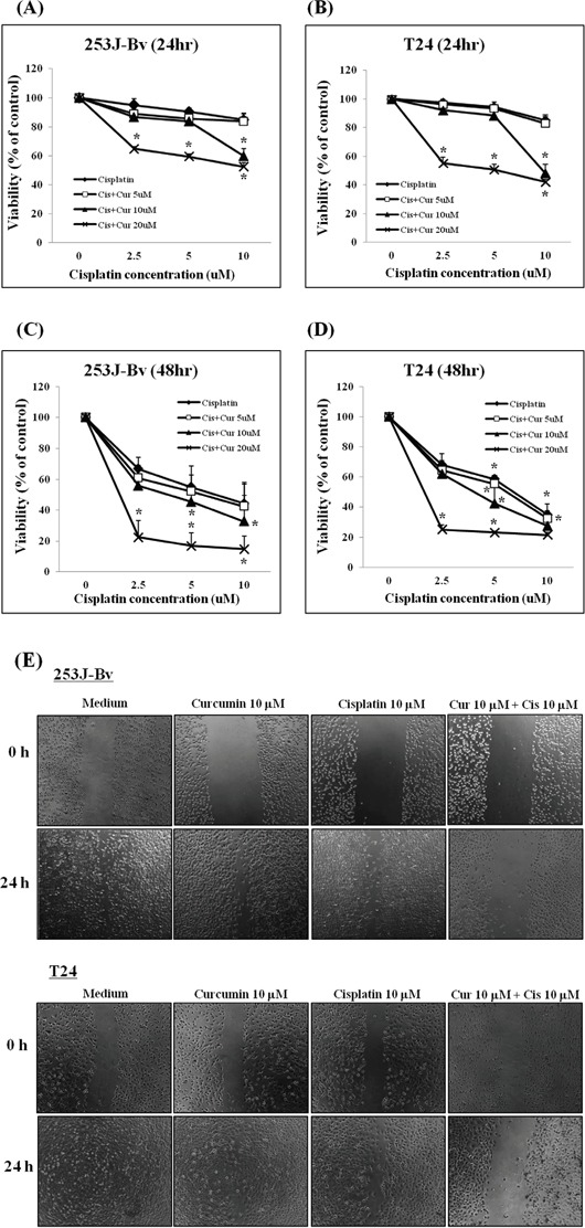
Figure 1. Proliferation rates of 253J-Bv and T24 cells after treatment with various cisplatin or curcumin concentrations.

A-D. Human bladder cancer cell lines (253J-Bv and T24) were treated with curcumin (5, 10, or 20 μM) and cisplatin (2.5, 5, or 10 μM) for 24 and 48 h. Cancer cell viability was measured by MTT assay. Data are expressed as the mean ± SEM of three independent experiments. *p <0.005 compared with medium alone was considered statistically significant. E. 253J-Bv and T24 cell monolayers were carefully scratched with a pipette tip and subsequently incubated with cisplatin (10 μM) and curcumin (10 μM) for 24 h. No treatment was administered to the control cancer cells. Migrating cells were photographed at 0 and 24 h post-wounding under a phase contrast microscope. The images represent three experiments showing similar results.
