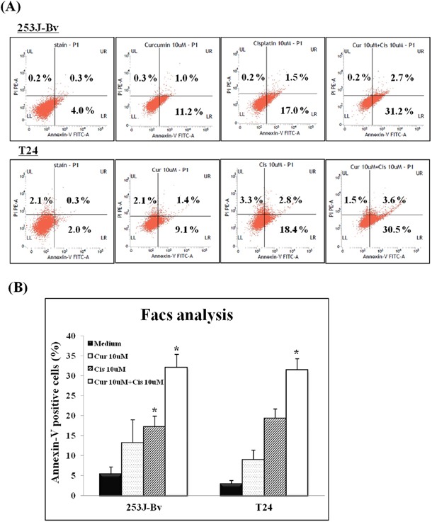
Figure 2. Detection of apoptotic cells in 253J-Bv and T24 bladder cancer cell cultures using flow cytometry after annexin V-FITC/PI staining.

253J-Bv and T24 cells were incubated for 24 h at 37°C with or without 10 μM curcumin and 10 μM cisplatin. After incubation, cancer cells were stained with PI and FITC-conjugated annexin V for flow cytometry. The diagram shows binding of annexin V (FL1) and PI in bladder cancer cells incubated for 24h with or without curcumin and cisplatin. The percentage of bladder cancer cells stained with both PI and FITC-labeled annexin V in each sample is indicated. Data are expressed as the mean ± SEM of three independent experiments. Significant differences from the results obtained for cells incubated with only medium are shown. *p <0.005 compared with medium alone was considered statistically significant.
