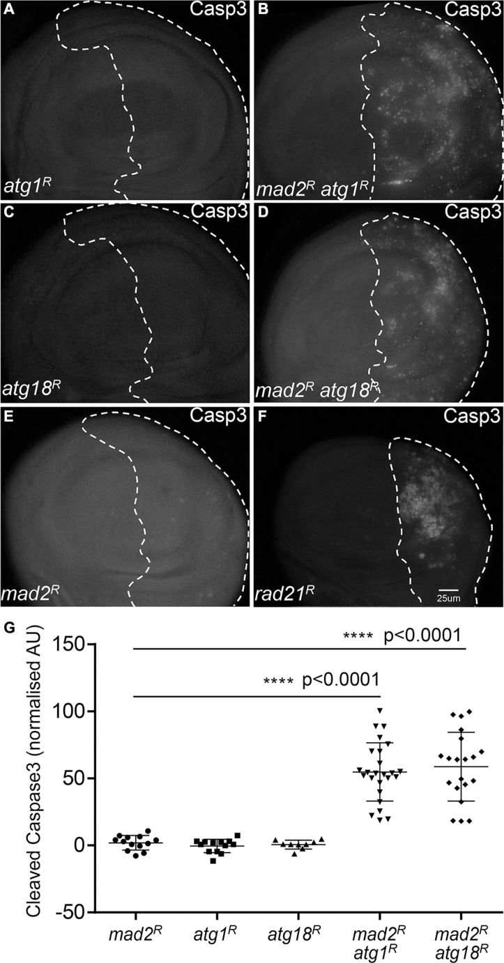
Figure 3. Blocking autophagy increases cell death in CIN cells.
Anti-cleaved caspase3 antibody staining was used to show the level of apoptosis. The indicated genes were knocked down in the posterior half of each wing disc as indicated by the dotted line and the rest of each disc was wild type. Knocking down either Atg1 ((A) engrailed > Gal4, UAS-CD8-GFP, UAS-Atg1RNAi) or Atg18 ((C) engrailed > Gal4, UAS-CD8-GFP, UAS-Atg18RNAi) did not cause apoptosis in these proliferating cells. However, knocking down Atg1 ((B) engrailed > Gal4, UAS-CD8-GFP, UAS-mad2RNAi, UAS-atg1RNAi) or Atg18 ((D) engrailed > Gal4, UAS-CD8-GFP, UAS-mad2RNAi, UAS-Atg18RNAi) in CIN cells, significantly increased the level of apoptosis in these cells relative to the CIN alone control (B, engrailed > Gal4, UAS-CD8-GFP, UAS-mad2RNAi). Depletion of rad21 ((F) engrailed > Gal4, UAS-CD8-GFP, UAS-rad21RNAi, UAS-Dicer2) shows for comparison the elevated apoptosis generated by a high CIN rate. Quantification of the cleaved caspase3 staining is shown in (G). In all cases n ≥ 9 and the error bars show 95% confidence intervals around the mean. The p values were calculated using two-tailed t-tests with Welch's correction.

