Figure 4.

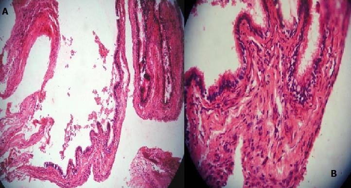
Photomicrograph showing a cystic lesion with papillary projections lined by pseudostratified columnar epithelium with some mucous pools and pseudoglandular areas (a) H&E stain at 10x magnification, (b) H&E stain at 40x magnification

Photomicrograph showing a cystic lesion with papillary projections lined by pseudostratified columnar epithelium with some mucous pools and pseudoglandular areas (a) H&E stain at 10x magnification, (b) H&E stain at 40x magnification