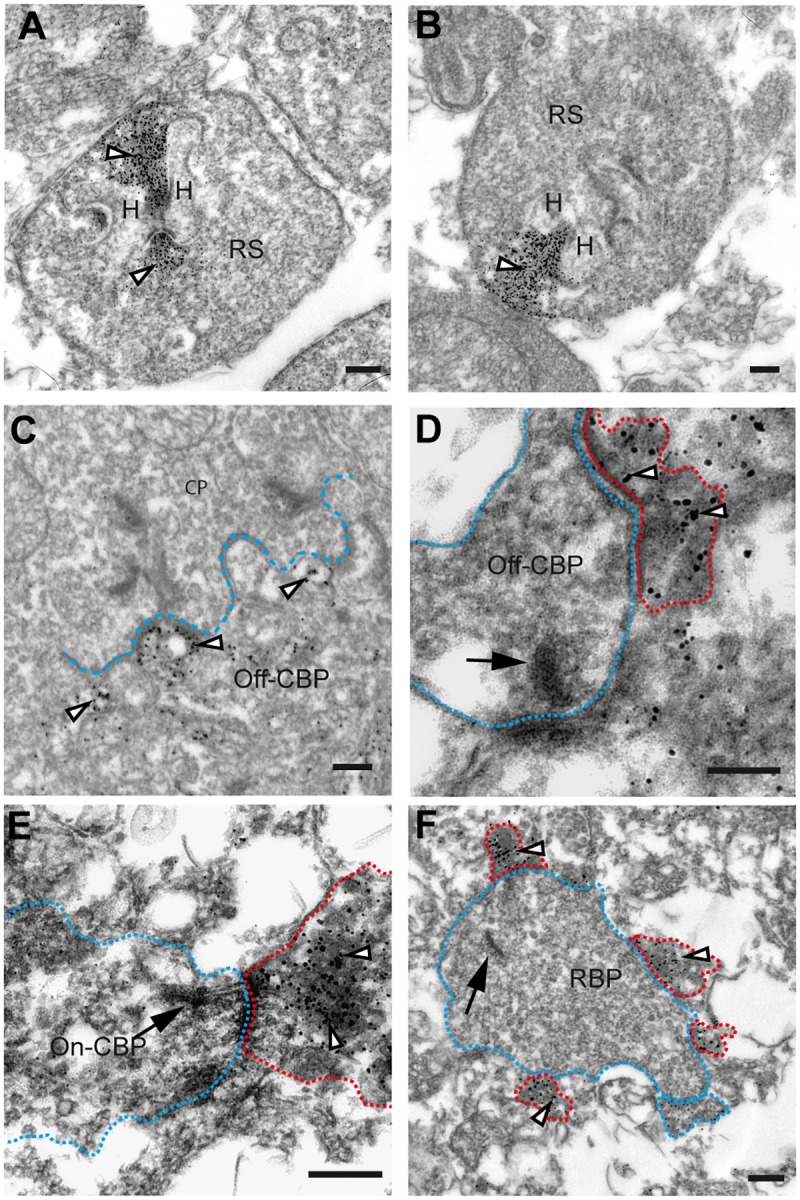
Fig 2. Ultrastructural localization of GluK5 in the outer and inner plexiform layer of the mouse retina.

(A and B) Electron micrograph of rod photoreceptor ribbons showing the ultrastructural localization of GluK5 immunoreactivity in rod spherules. Note the distribution of GluK5 immunoreactivity at the rod photoreceptor ribbon synapse covering the entire presynaptic ribbon structure (arrowheads). (C) Electron micrograph of a cone pedicle in the OPL showing GluK5 localization (arrowheads) in processes of OFF-cone bipolar cells. (D—F) Electron microscopic analysis of GluK5 distribution in the IPL. In the outer part of the inner plexiform layer GluK5 labeling is found in processes postsynaptic to OFF-cone bipolar cells (D). In the inner part of the inner plexiform layer labeling is seen in processes postsynaptic to ON-cone bipolar cells (E) and to rod bipolar cells (F). The arrows point to the presynaptic ribbon, the presynaptic terminals are surrounded with red dotted lines; the postsynaptic GluK5 positive elements are visualized with blue dotted lines. For better visualization in the electron micrographs presynaptic elements are visualized with blue and postsynaptic elements with red dotted lines. H = horizontal cell, RS = rod spherule, CP = cone pedicle, Off-CBP = off-cone bipolar cell, On-CBP = on-cone bipolar cell, RBP = rod bipolar cell. All scale bars: 100 nm.
