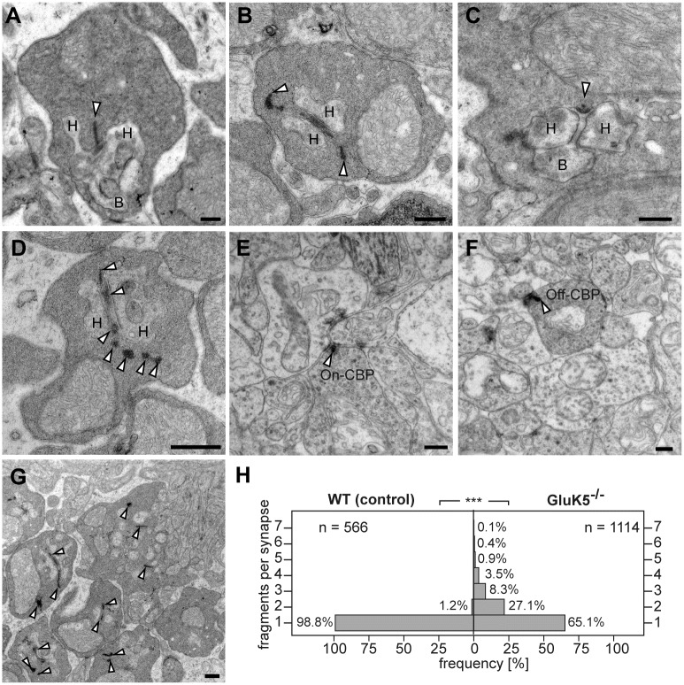
Fig 3. The absence of GluK5 disrupts the normal organization of presynaptic rod photoreceptor ribbons.
The electron micrographs show sections through rod ribbon synapses in the outer plexiform layer of wild-type (A) and GluK5-knockout retinae (B-D, G). Wild-type terminals (A) show normal synaptic architecture with a synaptic ribbon anchored to the presynaptic membrane and with postsynaptic elements formed by processes of horizontal and bipolar cells. (B-D) Rod terminals in adult GluK5-/- mice showing “disintegrated” synaptic ribbons. In some cases ribbon material seems to be free-floating within the rod terminal (B and D, arrowheads). (E –F) The synaptic ribbon structure is not affected at synapses in the IPL as demonstrated for ribbons of ON-cone (E) and OFF-cone (F) bipolar cells. The lower magnification (G) shows that most of rod photoreceptor ribbons are affected in the GluK5 knockout retina. (H) Histogram showing the number of ribbon fragments at rod photoreceptor terminals in GluK5 knockout and wild type animals. n = number of presynaptic elements analysed. ***P < 0,001 Mann-Whitney-U-Test. H = horizontal cell, B = Bipolar cell, Off-CBP = off-cone bipolar cell, On-CBP = on-cone bipolar cell. All scale bars: 250 nm.

