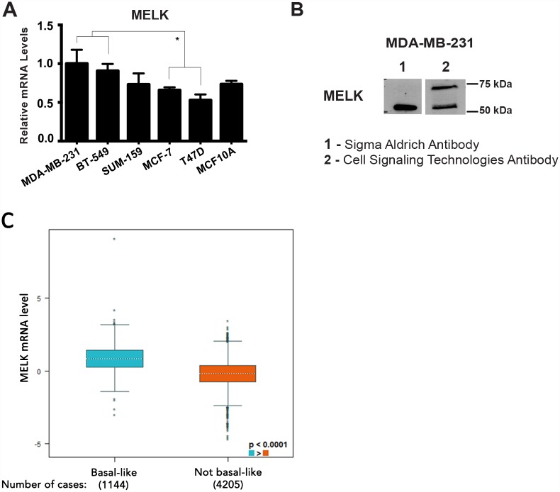
Fig 1. MELK is increased in TNBC cell lines and clinical samples.
(A) MELK is upregulated in the TNBC cell lines MDA-MB-231 and BT-549 compared to the Luminal A subtypes MCF-7 and T47D. (B) Western blot using two different MELK antibodies. In lysates from MDA-MB-231 cells, both the Sigma Aldrich and Cell Signaling Technologies antibodies detect a band near 52 kDa, but only the latter antibody detects the proposed full-length MELK isoform near 74 kDa. (C) MELK expression in basal-like and/or TNBC tumors (n = 1,144) compared with not basal-like tumors (n = 4,205). MELK mRNA level showed a significant increase in basal-like/TNBC tumors, p-value (Welch's) < 0.0001.

