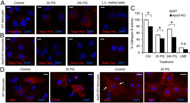
Fig 10. ApoD prevents lysosomal membrane permeabilization upon oxidative stress.
A-B. Cathepsin B activity monitored by Magic Red assay. The punctate fluorescent signal is proportional to its proteolytic activity, taking place in lysosomes. WT (A) and ApoD-KO (B) primary astrocytes are compared. L-leucyl-L-leucine methyl ester (LLME) is used as positive control for lysosomal membrane rupture. C. Cathepsin B activity is plotted normalized to values obtained in control WT astrocytes. PQ provokes a reduction of Cathepsin B activity that is recovered after 24 h treatment in WT astrocytes. Lack of ApoD results in reduced basal activity and unrecoverable activity loss after PQ insult. D. Representative fluorescence microscopy images of Galectin-3 signal in WT and ApoD-KO astrocytes under control and 2 h of PQ treatment. A switch from cytoplasmic to vesicular (lysosomal) Galectin-3 labeling occurs under oxidative stress conditions in WT astrocytes. The lysosomal labeling of Galectin-3 is evident for ApoD-KO primary astrocytes in control conditions (arrows), and increases under PQ treatment. Statistical differences in C were assessed by two-way ANOVA (p<0.001), and Holm-Sidak post-hoc method (p<0.001). Calibration bars: 20 μm (A), 10 μm (B).

