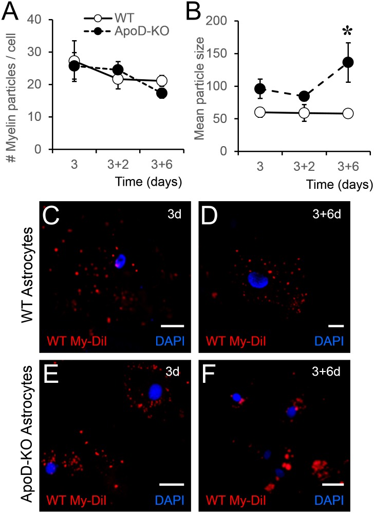
Fig 11. ApoD presence in mouse astrocytes is required for adequate processing of phagocytosed myelin.
WT and ApoD-KO astrocytes were exposed to DiI labeled myelin for 3 days, and DiI signal was evaluated by fluorescence microscopy 2 and 6 days after removal of myelin. Three-way ANOVA was used to evaluate variable interactions, followed by two-way ANOVA to detect the origin of differences. A. Number of DiI-myelin particles phagocytosed by primary WT and ApoD-KO astrocytes. No differences are found between genotypes, indicating comparable initial levels of phagocytic activity. B. Mean particle size of DiI-positive objects is dependent on time of treatment (p = 0.032, three-way ANOVA). The values at 6 days post-myelin removal account for the difference (p<0.001, Holm-Sidak method). Only ApoD-KO astrocytes show a significant increase in large myelin particles. C-F. Representative images of DiI-myelin signal in primary WT or ApoD-KO astrocytes after myelin exposure (3d; C,E) and 6 days after myelin removal (3+6d; D,F). Calibration bars: 20 μm.

