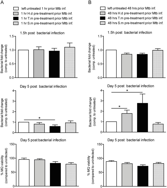
Fig 6. Mimicking chronic helminth infection by long-term antigen exposure results in lost hMDM control of Mtb.
hMDMs were treated with helminth antigens from H. diminuta (H.d; 1.5 μg/ml), T. muris (T.m; 1.5 μg/ml), or S. mansoni soluble egg antigen (S.m; 1.5 μg/ml) for 1h (A) or 48h (B) prior to infection for 1.5h with luciferase expressing Mtb (MOI = 2). Extracellular bacteria were washed away and Mtb phagocytosis was evaluated (upper horizontal panels), or washed hMDMs were incubated for 5 days at 37°C before the bacterial fold change was determined (middle horizontal panels). hMDMs pretreated with antigens for 1h prior Mtb infection had continuous presence of antigens throughout the experiment (A), whereas hMDMs pretreated for 48h received antigens prior Mtb infection only (B). A and B show the total bacteria (combined luciferase signal from hMDM lysate and supernatant) compared to untreated (only Mtb), data expressed as means ± SEM from 4 independent hMDM donors. Bottom horizontal panel, show hMDM viability after 5 days post Mtb infection measured using calcein AM uptake before lysates were generated for middle horizontal panels. Viability data are normalized against uninfected hMDMs set to 100%. p*<0.05 using One-way ANOVA.

