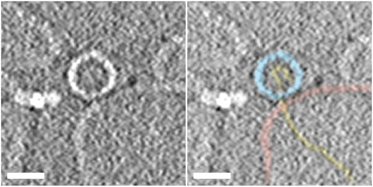
Fig 1. Section through a subtomograms from a cryoelectron tomographic reconstruction of a warmed virus-receptor- liposome complex showing RNA being translocated across the liposome membrane.
The samples were produced by heating virus-receptor-liposome complexes at 37°C for 4 min, mixed with colloidal gold, placed on carbon-coated Quantifoil holey grids and flash frozen, and cryo tomographic data were acquired and processed as in [22] The central section through a representative subtomogram containing a single complex from this data set is presented to summarise the path of the viral RNA from the interior of the virus, across the liposome membrane, and into the lumen of the liposomes during uncoating. The left panel shows a section through raw averaged subtomogram showing a virus particle (center) attached to a liposome (bottom right), with density for the RNA clearly extending from the middle of the particle across the membrane and into the lumen of the liposome. The bright feature to the left of the virus is a colloidal gold particle. The right panel shows the same section of the tomogram segmented to highlight the virus capsid (light-blue), the membrane bilayer (pink), and the RNA (gold). The scale bar in both panels is 25 nm.

