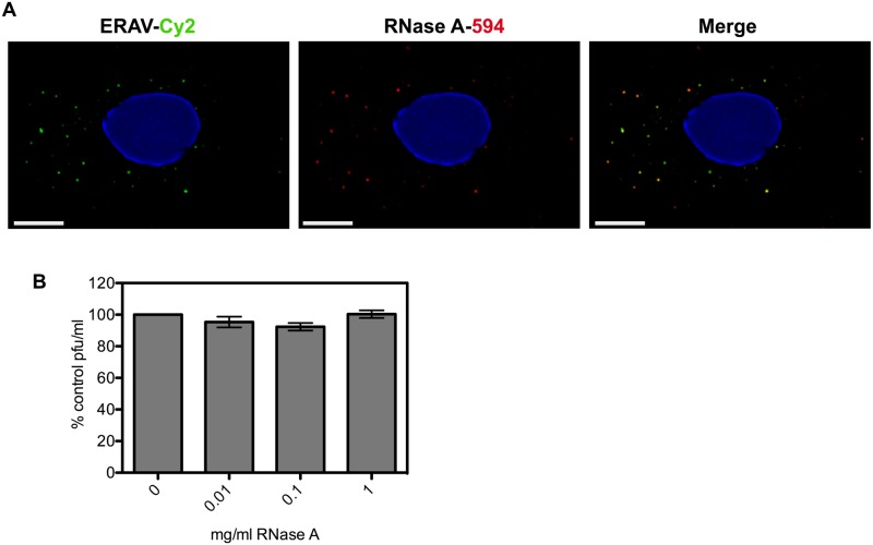
Fig 5. ERAV is co-internalized with RNase A but infectivity is not compromised.
A) Representative images of HeLa Ohio cells infected with ERAV conjugated to Cy2 (green, left panel) and RNase A-DyLight594 (red, middle panel) fixed 15 min post infection. The degree of co-internalization (Merge, right panel) was measured for 10 random cells (R = 0.86 +/- 0.09 (SD). Nuclei were stained with Hoechst (blue). Scale bar is 5 μm. B) Plaque assay of ERAV in the presence of 0–1 mg/ml RNase A. Plaque forming units (pfu) were expressed as percentage of no RNase A control. Data are from three independent experiments with error bars showing standard error.

