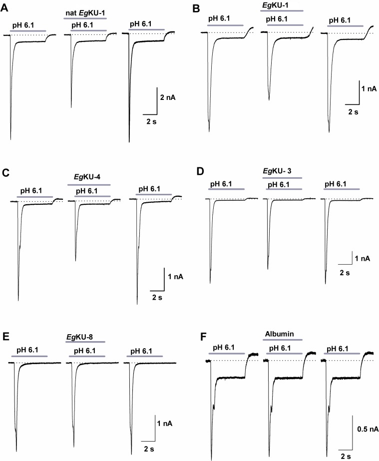
Fig 7. Inhibition studies with EgKU-1 and EgKU-4: results for ASIC currents from DRG neurons.
(A-C) Representative traces showing the acid (pH 6.1, 5 s) activated current under control conditions (left), after sustained (25 s) perfusion of 30 nM of each EgKU (center) and after 1 min washout of the inhibitors (right). Note that EgKU-1 and EgKU-4 reduced the amplitude of the Na+ current, that recombinant EgKU-1 reproduced the effect of the native inhibitor and that the recovery after washout was higher than 90% in all cases. (D-E) Representative traces from analogous assays with 30 nM of EgKU-3 and EgKU-8. The slight decrement of the current amplitude induced by EgKU-3 was significant (see the text for further details); EgKU-8 had no effect. (F) Albumin (15 μM) was used as negative control. Calibration in each case applies to the control, effect and washout recordings of each panel.

