. 2017 Feb 7;6:e23777. doi: 10.7554/eLife.23777

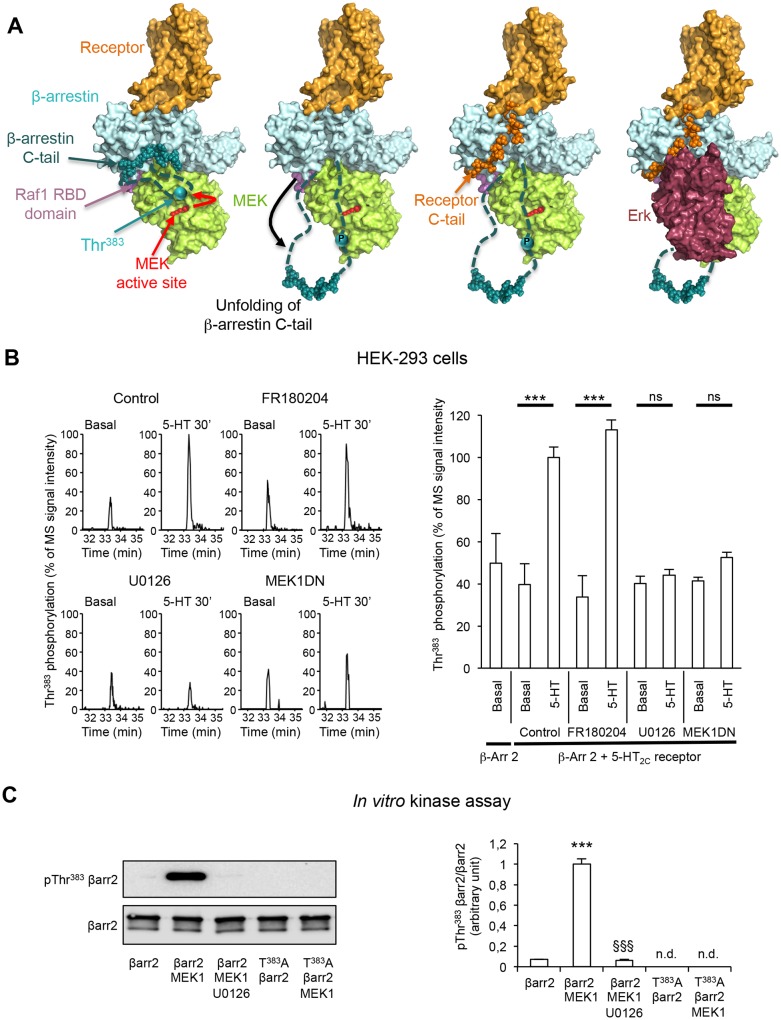
Figure 2. Role of MEK in the phosphorylation of β-arrestin2 at Thr383 elicited by 5-HT2C receptor stimulation.

(A) Mechanistic model of assembly of the 5-HT2C receptor/β-arrestin2/Erk module. Color code: receptor in orange, MEK in green, β-arrestin2 core in pale cyan and C-tail in cyan (the regions 351–384 and 394–419, which are not visible in 3D structure are represented by dashed lines, the region 385–393 is represented by spheres), Erk in dark red, Raf-1 RBD domain in pink. In this model, we hypothesize that Thr383 phosphorylation by MEK takes place within the assembled receptor/β-arrestin/Raf/MEK complex and results in a movement of β-arrestin2 unfolded 350–393 segment away from the first β-strand of β-arrestin, leaving space for further interaction with the receptor C-terminal domain (orange spheres) and recruitment of Erk, and its subsequent phosphorylation by MEK. For the clarity of the figure, the extremity of the β-arrestin C-tail is represented by spheres even in its unfolded state, although the real 3D structure is unknown. (B) Representative extracted ion chromatograms of the peptide in cells expressing 5-HT2C receptor, pretreated with either vehicle (control) or FR180204 (10 µM for 18 hr) or U0126 (5 µM for 30 min) or coexpressing MEK1 dominant-negative mutant (MEK1DN), and challenged with vehicle (Basal) or 5-HT (1 µM) for 30 min. The histogram represents the means ± SEM of the corresponding ion signal intensities (normalized to values in 5-HT-stimulated cells in Control condition) obtained in three independent experiments. One-way ANOVA: F(8,18) = 15.69, p<0.0001. ***p<0.001 vs. corresponding basal value. (C) YFP-tagged β-arrestin2 (wild-type or Thr383Ala mutant) purified from transfected HEK-293 cells was incubated with active MEK1 for 15 min at 37°C. When indicated, U0126 (5 µM) was included in the incubation medium. Thr383 phosphorylation was assessed by sequential immunoblotting with the antibody raised against phospho-Thr383 β-arrestin2 and the anti-β-arrestin2 antibody. Means ± SEM of results from four independent experiments are shown on the histogram. n.d.: not detectable. One-way ANOVA: F(2,9) = 352.2, p<0.0001. ***p<0.001 vs. immunoreactive signal in absence of MEK; §§§ p<0.001 vs. corresponding condition in absence of U0126.

DOI: http://dx.doi.org/10.7554/eLife.23777.010

Figure 2—source data 1. This file contains raw values used to build Figure 2B, C.
DOI: 10.7554/eLife.23777.011

Figure 2.

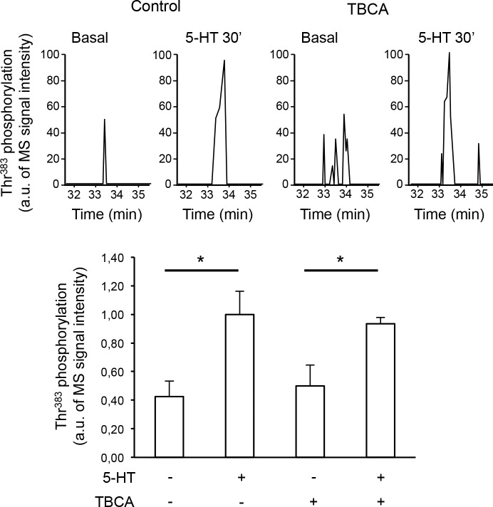
Figure 2—figure supplement 1. Thr383 phosphorylation is not mediated by casein kinase 2.

Figure 2—figure supplement 1.

(A) Representative extracted ion chromatograms of the EIDIPVDTNLIEFDTNYAp383TDDDIVFEDFAR peptide from YFP-tagged β-arrestin2 in cells expressing 5-HT2C receptor and challenged with vehicle (Basal) or 1 µM 5-HT for 30 min in absence or presence of TBCA (1 µM, added 15 min before the onset of 5-HT treatment). Two other independent experiments performed on different sets of cultured cells yielded similar results. (B) The histogram represents the means ± SEM of ion signal intensities of the peptide (normalized to values in 5-HT-stimulated cells) obtained in the three experiments. One-way ANOVA F(3,8) = 6.762, p=0.013. *p<0.05 vs. corresponding vehicle.
Figure 2—figure supplement 1—source data 1. This file contains raw values used to build Figure 2—figure supplement 1.
DOI: 10.7554/eLife.23777.013
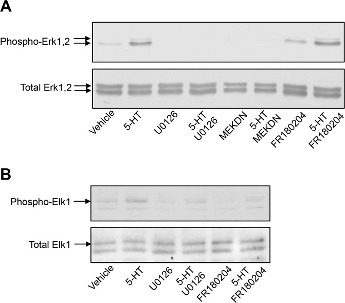
Figure 2—figure supplement 2. Impact of MEK and Erk1/2 pharmacological inhibitors and of co-expression of MEK dominant-negative mutant on 5-HT2C receptor-operated Erk1/2 and Elk1 phosphorylation.

Figure 2—figure supplement 2.

(A) Cells expressing 5-HT2C receptor were pretreated with vehicle or U0126 (5 µM for 30 min) or FR180204 (10 µM for 18 hr) and then exposed to vehicle or 5-HT (1 µM) for 5 min. When indicated, a dominant-negative mutant of MEK (MEKDN) was co-expressed by 5-HT2C receptor. Erk1/2 activation was assessed by sequential immunoblotting with the antibody recognizing phospho-Thr202/Tyr204-Erk1/2 and total Erk1/2. Immunoblots representative of three independent experiments are illustrated. Note that FR180184 did not affect 5-HT2C receptor-operated Erk1/2 phosphorylation, indicating that it does not inhibit MEK. (B) The impact of MEK and Erk1/2 inhibitors on phosphorylation of the Erk1/2 substrate Elk1 was assessed by sequential immunoblotting with the antibody recognizing phospho-Elk1 and total Elk1. Immunoblots representative of two independent experiments are illustrated.
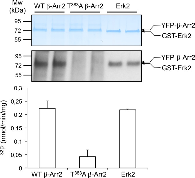
Figure 2—figure supplement 3. In vitro phosphorylation of β-arrestin2 versus Erk2 by MEK1.

Figure 2—figure supplement 3.

YFP-tagged β-arrestin2 (wild-type or Thr383Ala mutant) purified from transfected HEK-293 cells or purified non-activated Erk2 (~1 µg each) were incubated with active MEK1 in presence of [γ-32P]-ATP (2 µCi/nmol) for 10 min at 37°C. Proteins were separated by SDS-PAGE and stained with Coomassie colloidal blue (top image) and 32P incorporation into the different substrates was monitored by autoradiography (bottom image). The data in the histogram, expressed in nmol/min/mg enzyme, represent the means ± SD of 32P incorporation into β-arrestin2 and Erk2 protein bands in the corresponding experiment after radioactive background subtraction for each lane.
Figure 2—figure supplement 3—source data 1. This file contains raw values used to build Figure 2—figure supplement 3.
DOI: 10.7554/eLife.23777.016