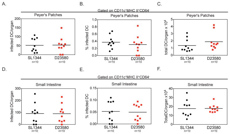
Figure 3. Similar levels of infected DC are present in gut tissues from mice infected with ST19 or ST313 S. Typhimurium isolates.
(A–I) Streptomycin-pretreated C57BL/6 mice were orally infected and mice were sacrificed 2 dpi. Single cell suspensions of the Peyer’s patches and small intestine were stained for cell surface markers and intracellular Salmonella and analyzed by flow cytometry. The total number of infected DCs per organ (A, D), percent of DCs infected (B, E,) and total number of DCs per organ (C, F) are presented for Peyer’s patches (A–C) and small intestine (D–F). Data are combined from 2 independent experiments with 5 mice per group. (A–F) The mean for each group is shown and statistical significance was determined by the Mann-Whitney test. * P≤0.05. See also Figure S1 and S4.

