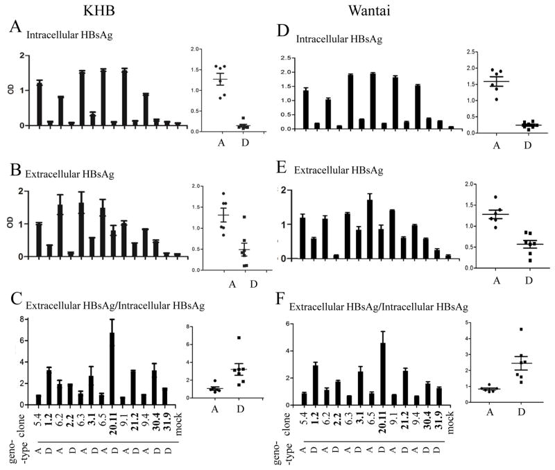
Fig 1.
Comparison of intracellular and extracellular HBsAg from Huh7 cells transfected with dimeric constructs of genotype A and genotype D. SphI dimers of 6 genotype A clones and 7 genotype D clones were transiently transfected to Huh7 cells, followed by ELISA detection of intracellular HBsAg (diluted 1:1600 for KHB kit, 1:800 for Wantai kit) and secreted HBsAg (diluted 1:800 for KHB kit, 1:400 for Wantai kit) 5 days posttransfection. Furthermore, the ratio of extracellular HBsAg/intracellular HBsAg was calculated (C and F). The ELISA data were based on 4 independent transfection experiments.

