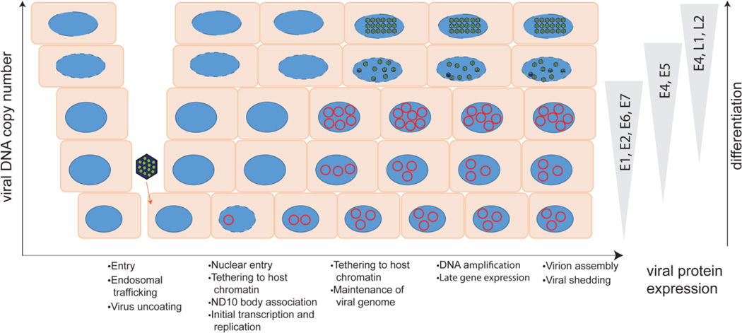
Figure 2. Overview of the viral infectious cycle.
The virus accesses the basal keratinocytes through a microabrasion. After entering the cell, the virus traffics through the endosome. Breakdown of the nuclear envelope during mitosis allows the virus to enter the nucleus and viral DNA is observed on mitotic chromosomes, in complex with the L2 protein. The L2 genome complex then localizes to ND10 bodies and early gene expression occurs. After a short burst of replication, the genome is maintained at a low copy number in the dividing cells in the lower levels of the epithelium. As the infected keratinocytes differentiate, the genome is amplified to high levels and late genes are expressed. The viral genome is assembled in capsids in the superficial layers of the epithelium, and viruses are shed from the surface in viral-laden squames. The different steps in the viral life cycle are listed below the diagram; host restriction factors can interfere at many stages of infection.

