Figure 1.
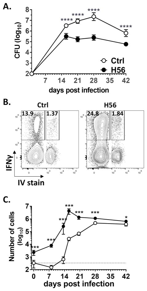
H56/CAF01-mediated protection is accompanied by accelerated recruitment of Mtb-specific T cells to the lung parenchyma.
CB6F1 mice were vaccinated and rested 6 weeks prior to aerosol Mtb challenge. (A) Total Lung CFU from adjuvant-only control (Ctrl, ○) and H56/CAF01-vaccinated (H56, ●) CB6F1 mice. Symbol, mean±SD of 5–6 mice. (B) Mice were stained i.v. with anti-CD45-FITC to distinguish parenchymal (IV−) from vasculature (IV+) cells, followed by ex vivo stimulation of lung cells with H56 protein and ICS staining for IFNγ/TNFα/IL-2/IL-17 production to enumerate H56-specific CD4 T cells. H56-specific IFNγ production by IV+ and IV− CD4 lung cells from a control (left) and a H56/CAF01-vaccaninated (right) mouse 3 weeks after aerosol Mtb infection. (C) Total number of IV− H56-specific CD4 lung cells from adjuvant only control (○) and H56/CAF01-vaccinated (●) CB6F1 mice before and after aerosol Mtb infection, based on ICS staining for cells producing any cytokine (IFNγ, TNFα, IL-2, or IL-17). Data are pooled from 3 independent experiments each with 3–6 mice per timepoint. Symbol, mean±SEM. Dotted line represents assay background, i.e. mean pre-infection H56-specific response in adjuvant-only control mice. *p<0.05,**p<0.01, ***p<0.001 ****p<0.001 vs control by unpaired T test.

