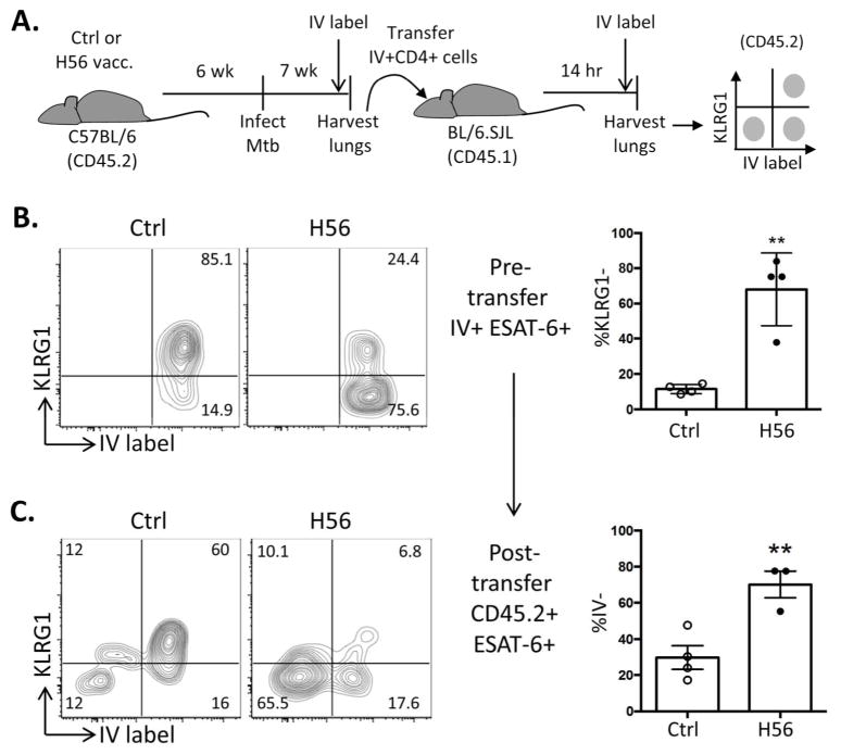
Figure 5.
Vaccine-specific Lung Vasculature CD4 T cells from H56/CAF01-vaccinated mice efficiently traffic into the Mtb-infected Lung parenchyma\r\n (A) Schematic of adoptive transfer system to study the parenchymal homing ability of lung-circulating H56/CAF01-induced CD4 T cells. Total IV+ lung CD4 T cells were FACS-sorted from 7wk Mtb-infected adjuvant control or H56/CAF01-immunized BL/6 mice (CD45.2) and transferred into infection-matched congenic recipients (CD45.1). After 14h, the migration of donor cells into the lung parenchyma was determined by a second i.v. stain of recipients prior to lung cell isolation and surface staining. (B) KLRG1 expression of ESAT-64-17 tet+ cells from sorted IV+ donor CD4 T cells from individual adjuvant control (left) or H56/CAF01-vaccinated (right) mice prior to transfer. Bar, mean±SD of 4 mice. (C) KLRG1 expression and IV-labeling of CD45.2+ I-Ab:ESAT-64-17 tetramer + donor CD4 T cells isolated from the lungs of recipient mice to assess trafficking and phenotype of transferred cells. Bar, mean±SD of 3–4 mice. Data representative of 2 experiments with similar results. * p< 0.05, **p<0.01 by unpaired t-test.\r\n

