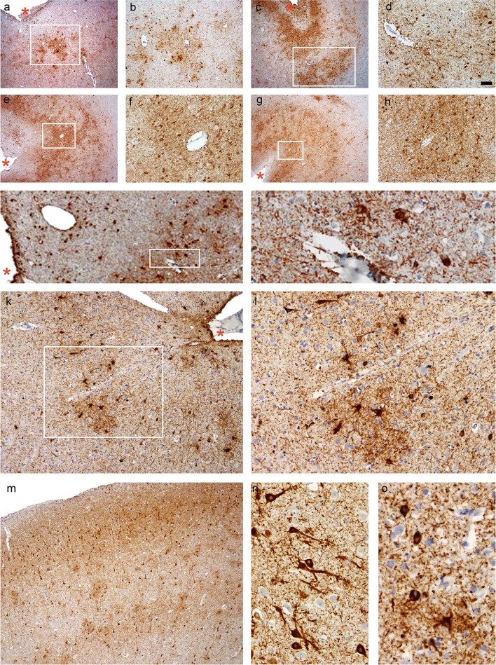
Fig. 2.
CTE pathology. a, b Case 1, parietal cortex, c, d Case 2, temporal cortex, e, f Case 5, temporal cortex, g, h Case 5, posterior frontal cortex (including the motor cortex), and i–l Case 6, temporal cortex. b, d, f, h, j, and l are images at high magnifications of the boxed regions on (a), (c), (e), (g), (i), and (k), respectively. a–l Patchy tau aggregates in neurons, astrocytes, and cell processes found preferentially at the depths of the cortical sulci with multiple perivascular foci. Cortical sulci are marked by asterisks. m Neuronal tau aggregates preferentially affecting superficial cortical layers (layers II–III) in CTE (Case 6, temporal cortex), which contrasts with the involvement of the deep cortical layers in Alzheimer’s disease (see Fig. 3). n Prominent proximal dendritic swellings in CA4 hippocampal subregion (Case 5). o Dot-like structures in the neuropils (Case 6, temporal cortex). All sections immunostained for AT8. Bar represents 100 µm in (a), (c), (e), (g), (i), and (m), 40 µm in (b), (j), and (k), 20 µm in (d), (f), (h), (l), and (n), and 10 µm in (o)

