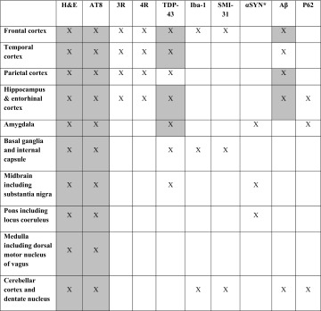
Table 1.
Brain regions evaluated in the six post-mortem cases
Grey boxes represent the sampling regions recommended by the preliminary NINDS criteria for the neuropathological diagnosis of CTE [30]
H&E haematoxylin and eosin; antibodies for immunohistochemistry, 3R 3-repeat tau, 4R 4-repeat tau, αSYN alpha-synuclein, Aβ beta-amyloid, AT8 tau, Iba-1 microglia, p62 for argyrophilic grains in amygdala and hippocampus and C9orf72 inclusions in hippocampus and amygdala, SMI-31 phosphorylated neurofilament, TDP-43 transactive response DNA-binding protein, 43 kDa
aIf αSYN is positive in midbrain, pons and amygdala, then including frontal and temporal cortices and hippocampus

