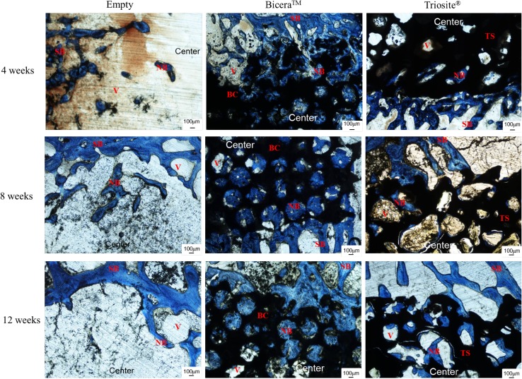
Fig. 3.
Histological image of rabbit femoral condyle tissue section. In Bicera™ group, new bone (blue area) grew into porous structure of bone substitute (black area). In Triosite™ group, there was an obvious interface between bone substitute (black area) and host bone (blue area) (SB surrounding bone, NB new bone, BC Bicera™, TS Triosite™, Center central site in defect)

